Advanced Imaging
In Room 161 of Rockwell Integrated Sciences Center sits a versatile, multipurpose machine that puts high-resolution imaging and data collection directly at the fingertips of faculty and students. Situated on top of a large pedestal, it emits a soft hum and somewhat formidable presence even when it isn’t in use. Multiple screens—including one on the wall for multi-student observation—allow for data-rich imaging and both wet and dry samples to be examined.
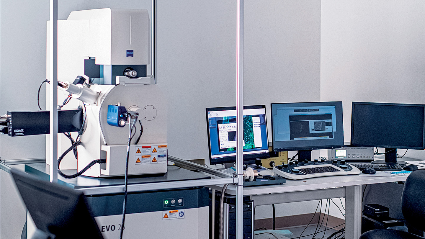
The instruments of the EVO combine high-performance scanning electron microscopy with an intuitive, user-friendly experience that appeals to both professors, who have experience as microscopists, and new users, such as students who are still training. The Dyer Center contributed funding to support the installation the SEM.
In the scientific community, the machine is known as a scanning electron microscope (SEM), which uses a beam of electrons, instead of light, to see an image. The exact model of the microscope is a Zeiss EVO 25. To the Lafayette community, it’s a high-tech, industry-standard, long-awaited tool that will open up numerous research and collaborative opportunities with other institutions.
It has been one year since the National Science Foundation awarded Lafayette a grant to acquire the SEM, with installation originally scheduled to happen in 2020. The sophisticated piece of equipment is a rare acquisition for an undergraduate college. Not only will it aid in research and hands-on teaching and learning for Lafayette’s biology, chemical and biomechanical engineering, civil engineering, environmental science, geology, neuroscience, and physics faculty and students, but outside parties will be encouraged to utilize the SEM for high-level collaborative research and educational opportunities.
The sophisticated piece of equipment is a rare acquisition
for an undergraduate college.
There are already plans in place to have a class visit from Phillipsburg High School, and faculty from Moravian University also will be making use of the microscope.
“Zeiss undoubtedly makes the best microscopes in the world. The SEM complements a light microscope that we already have upstairs [in Rockwell], and you can take the same specimen and look at it on both microscopes and correlate the two images,” shares James Dearworth, associate professor of biology, who wrote the grant to obtain the SEM. “We’re ultimately going to use it as a tool of outreach. We put a lot of thought into figuring out where this thing is going to live and sharing how the system works so that it goes beyond Lafayette.”
Among other components of what makes the SEM special, it features class-leading X-ray spectroscopy and cathodoluminescence for sample analysis, introduces research-grade nitrogen gas around delicate specimens so that they can be examined, and rests on a plinth that shelters the SEM from environmental vibrations that might interfere with the imaging process.
Here is how faculty members plan to utilize the cutting-edge technology they now have at their fingertips:
James Dearworth
associate professor of biology
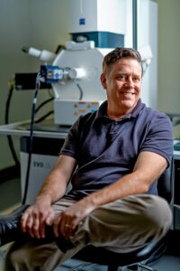
James Dearworth, associate professor of biology, wrote the grant to obtain the EVO microscope.
“I do anatomy, so I look at tissues—including tissues of the eye—and I was very interested in using this to look at the anatomy of turtles. But for the biology students, we’ve designed an exercise where they’re going to look at pollen in plants. They’re going to go out into the field, collect these species, and then they’re going to compare pollen from them and see if they fit into two different types—gymnosperms and angiosperms. They’ll start by looking at pollen under the light microscope, and then they’re going to use the SEM to look at topology. The idea is, can they use the imaging to determine the differences?
“I’m going to work with (associate professor of biology) Megan Rothenberger ’02 and (general biology lab coordinator) John Drummond, and we’re going to design a simple investigation where the challenge will be balancing the number of samples. Then in the spring, we’re going to use [the SEM] to get a little higher resolution to identify viruses—phages from soil samples that have been collected as part of the second semester. After they’ve identified the sequences, they can actually take those images and see what the phages look like. The students will get to learn by doing, which is what I think Lafayette is all about.”
Tamara Carley
associate professor of geology
“My specialty is igneous petrology—the study of rocks that form from magma. I start out with my students thinking about features that are visible on the landscape. It’s my job to take what you see when you’re driving around the countryside, or hiking, down to smaller and smaller levels—from mountains, to rocks, to minerals—and then down to the chemistry building those minerals. Without an instrument like this [the EVO 25 SEM], there are a lot of leaps of faith that we have to make; this SEM will remove a lot of the mystery. My primary use for this instrument will include looking at the compositional zoning inside of minerals. As minerals grow, it’s sort of like looking at the inside of a tree, and you can see the earliest chapters of their history. I use that perspective to reconstruct stories of magma generation, evolution, and instability.
“There are chemical fingerprints that help you reconstruct that story along the way; you can see elements coming in and out of the magmatic system in response to temperature changing and pressure changing. A lot of that is invisible until you can get an instrument like this with a cathodoluminescence detector. For me, this is what really sets this instrument apart from others. I’m planning to use the EVO in my mineralogy, petrology, and geochemistry classes. In mineralogy, students will take sand collected over summer vacation and distill it down to get the major minerals in geologic history—taking teeny tiny little crystals that are barely visible under a standard microscope, and magnifying them thousands of times to see their physical and chemical features.”
Annemarie Exarhos
assistant professor of physics
“I use light and lasers to look at crystals and defects in problematic areas, and to characterize them and understand them. In particular, I look at the atomic scale effect. So, what happens if you take a diamond and replace one atom with nitrogen and remove the neighboring atom? It turns out, you get a really interesting effect; because it converts information into light, you can use it as the base of a quantum computer. That qubit is like a quantum analog computation, but it has different properties. We do this using confocal fluorescence microscopy. This means we shine light on it, and we detect light coming out of it from the single photons, which is the smallest amount of light we can possibly get.
“We know crystals that have really interesting and potentially useful defects like this for quantum computation or other types of quantum technologies. But there are many types of crystals and there are many types of defects, and a lot is really unknown. So, I’m looking at basic characterization of new types of effects and crystals that can be used for these quantum technologies. I have two intended classes that I want to use this for, and one of them is a 400-level solid-state physics, where we talk a lot about how to characterize crystals and what different crystalline structures give you in terms of the actual physical properties of the material. In order to understand that, you need to understand what the crystal structure is, what things look like. I’m hoping to bring students in to actually see this kind of electron backscatter diffraction, and compare it with X-ray diffraction. And then I also teach an introductory-level class for first-year students, which includes a section on nanoscience. I’m hoping to bring them over and put a sample in, and see what it looks like.”
Zoe Boekelheide
associate professor of physics
“In my research, I work on magnetic nanoparticles and study magnetic properties. If you want to magnetically characterize them, you also have to know what they look like structurally—otherwise, you’re just characterizing some unknown sample. The benefit of this instrument [the EVO 25 SEM] is it has much higher resolution than the previous SEM that we’ve had access to here. A lot of my nanoparticles are in that 30- to 50-nanometer range, and it will be extremely helpful to be able to see that. In magnetic nanoparticles, each little nanoparticle is basically a tiny magnet. I’m interested not just in the structure of individual nanoparticles, but how they form
a superstructure.
“My work with students is through the EXCEL program, and future students will be working with the SEM to characterizenew particles.”
Christopher Hawley
assistant professor of physics
“I work in the field of condensed matter physics. My summer students were generally starting to ramp up the growth of nanoscale crystals. So, a lot of what I do falls under the category of nanoscience and nanotechnology. We will be growing nanowires—growing thin films—and it’s really great to have the SEM so that we can directly image these things. They’re so small that when you put them under a microscope, you don’t see much that makes sense. They’re beyond what visible light can resolve in an optical microscope. So that’s really the beauty of having something like [the EVO 25] that can go thousands of times smaller than an optical microscope.
“You’re growing these nanowires from scratch. We start with gas vapors, and we’re literally building an atom layer by layer to grow these nanostructures. So, this is very integral to being able to tell that you’ve grown the right morphology and the right shape of nanostructure that you’re interested in. Not only that, but being able to tell something about the crystallography and the chemistry of the nanostructure you’ve grown helps to complete the picture.
“Annemarie and I are working with a joint collaborator in Philadelphia, and they look at everything from nanowires to small-scale transistors to electronics that will go into cellphones and computer processors. We’ll be able to take the optics data from that experiment and then bring the same sample over here, and take a map of the same circuitry and create a chemical map. There are probably only a dozen places in the world that do that well, so that’s really exciting. And to have our students be able to work on this here at Lafayette, which is super exciting for them.”
Daniel Proud
assistant professor of biology (Moravian University)
“I’m very excited to be able to collaborate with folks at Lafayette and be able to access this piece of equipment, which you don’t find at every institution. So, it’s really nice that it’s available, and it’s pretty exciting to be able to get students involved in research that they can make use of the SEM. Most of what I’ll be using it for is to take high-magnification images of these animals that I study—mostly arachnids—but it could possibly include some insects if we start doing things with some pollinators.
“I work specifically with daddy long-legs spiders. We dissect them and then look at them in parts. We’re interested in some of these very fine details, like sensory hairs, glandular openings, little spines that you find on the body, or tubercles you find in the body. We often dissect and look at the genitalia because those are species-specific, or tell us something about what family they belong to, or we can differentiate two species based on that. There are a lot of structures that we can look at using SEM that we can’t quite get enough detail using just standard microscopy.”
More Cutting-Edge Equipment Arrives on Campus
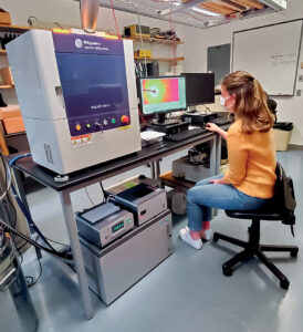 Earlier this semester, Lafayette’s Hugel Science Center became home to an XtaLAB mini II single crystal X-ray diffractometer, which provides structure analysis capabilities in a way that minimizes the possibility of misaligning or damaging the system. Acquisition of the diffractometer also ensured that students no longer have to travel outside to other institutions to access this rare and useful tool.
Faculty and students from chemistry, geology, physics, and environmental engineering are utilizing the XtaLAB mini II to determine their crystal structures.
Addition of the new equipment was made possible by a grant from the McCutchen Foundation, with a request for the equipment put in by Chip Nataro, Marshall R. Metzgar Professor and head of chemistry, and the grant itself submitted by Markus Dubischar, associate dean of the curriculum, whose academic interests include classical civilizations. The foundation provided $30,000 to support the diffractometer purchase.
The simplified design and easy-to-run software for the diffractometer make it the perfect teaching instrument for chemical crystallography, and students began to work with the instrument soon after its installation.